domingo, 8 de marzo de 2015

L15: ANIMAL CELLS vs PLANT CELLS


1. OBJECTIVES


- Identify the major components of cells.
- Differenciate between animal and plant cells,
- Measure dimensions of the entire cell and the nucleous.

2. MATERIALS


- Toothpick
- 2 Slides
- 2 Covers slips
- Distilled water
- Methylene blue
- Iodine
- Onion
- Glycerine
- Two whatch glasses
- Dropper
- Needle

3. PROCEDURE


- PLANT CELLS OBSERVATION:

1. Pour some distilled water into watch glass.
2. Peel off the leaf from half a piece of onion and using forceps, pull out a piece of transparent onion peel (epidermis) from the leaf.
3. Put the epidermis in the watch glass containing distilled water.
4. Take a few drops of iodine solution ( or safranin) in a dropper and transfer into another watch glass.
5. Using a brush ( or a needle), transfer the peel into the watch glass containing the dye, Let this remain in the safranin solution (or iodine) for 30 seconds, so that the peel is stained.
6. Take the peel from the iodine solution and place it in the watch glass containing distilled water.
7. Take a few drops of glycerine in a dropper and pour 2-3 drops at the center of a dry glass slide.
8. Using the brush, place the peel onto the slide containing glycerine.
9. Take a cover slip and place it gently on the peel with the aid of a needle.
10. Remove the extra glycerine using cellulose paper.
11. View it in microscope.

- CHEEK CELLS OBSERVATION:

1. Gently scrape the inner side of the cheek using a toothpick, wich will collect some cheek cells.
2. Place the cells on a glass slide that has water on it.
3. Mix the water and the cheek cells using a needle and spread them.
4. Dry the sample under the light to fix the sample on the slide,
5. Take a few drops of methylene blue solution using a dropper and add this to the mixture on the slide,
6. After 2-3 minutes remove any excess water and satin from the slide using cellulose paper.
7. Take a clean cover slip and lower it carefully on the mixture with the aid of a needle.
8. Using the top of the needle, press the cover slip gently to spread the epithetial cells,
9. Remove any extra liquid around the cover slip using cellulose paper.

4. RESULTS AND CONCLUSIONS


PLANT CELLS:

MRcell: 6,9/400= 0,017cm
0,17x10000= 172,5microns

MRnucleous; 400=0,7x10000/X
X=7000/400=17,5 microns


CHEEK CELLS:
400= 1,5x10000microns/X
X=1,5x10000microns/400=37,5

400=0,3x10000microns/X
X= 0,3x10000microns/400=7,5microns

lunes, 16 de febrero de 2015

L12: DNA EXTRACTION


1. INTRODUCTION


Deoxyribonucleic acid (DNA) is a nucleic acid that encodes the genetic instructions used in the development and functioning of all known living organisms and many viruses. Nucleic acid are biopolymers formed by simple units called nucleotides. Each nucleoide is composed of a nitrogen- containig nucleobase ( G,T,C,A) as well as a monosacharide and a phosphate group.
These nucleoides are joined to one another in a chain by covalent bonds between the sugar of one nucleoide and the phospate of the next. Most DNA molecules consist of two strands coiled around each other to form a doble helix. Hydrogen bonds bind the nitrogenous bases of the two separate strands.
The two strands run iin opposite directions to each other and are therefore anti-patallel. Moreover the bases of the two opposite strands unit according to base pairing: A-T and C-G.
Within cells, DNA is organized into structures called chromosomes.

2. OBJECTIVES


- Study DNA structures.
- Understand the process of extracting DNA from a tissue.

3. MATERIAL


- 1L Erlenmeyer flask
- 100mL beaker
- 10mL graduated cylinder
- Small funnel
- Glass stirring rod
- 10mL Pipet
- Knife
- Safety goggles
- Cheesecloth
- Kiwi
- Pineapple juice
- Distilled water
- 90% Ethanol ice-cold
- 7mL DNA buffer
- 50mL dish soap
- 15g NaCl
- 900mL tap water

4. PROCEDUCE


1. Peel the kiwi and chop it to small pieces. Place the pieces of the kiwi in one 600mL beaker and smash with a fork until it becomes a juice pure.

2. Add 8mL of buffer to the mortar.

3. Mash the kiwi puree carefully for 1 minute without creating many bubbles.

4. Filter the mixture: put the funnel on top of the graduated cylinder. Place the cheesecloth on top of the funnel.

5. Add beaker contain carefully on top of cheesecloth to fill the graduated cylinder. The juice will drain through the cheesecloth but the chucks of kiwi will not pass through in to the graduated cylinder.

6. Add the pineapple juice to the green juice ( you will need about 1mL of pineapple juice to 5mL of the green mixture DNA solution). This step will help us to obtain a purer solution of DNA . Pineapple juice contains an enzyme that breaks down the proteins.

7. Tilt the graduated cylinder and pour in an equal amount of ethanol with an automatic pipet. Put the ethanol through the sides of the graduated cylinder very carefully. You will need about equal volumes of DNA solution to ethanol.

 8. Place the graduated cylinder so that it is eye level. Using the stirring rod, collect DNA at the boundary of the ethanol and kiwi juice; only the stir in the above ethanol layer!

9. The DNA precipitate looks like long, white and thin fibers.

10. Gently remove the stirring rod and examine what the DNA looks like.


5. OBSERVATIONS/ CONCLUSION


We can observe the fibers of DNA.

6. QUESTIONS


1. What did the DNA looks like?

The DNA looks like white  and thin fibers.

2. Why do you mash the DNA? Where it is located inside the cells?

The crush to extract the liquid from the kiwi, the DNA is in the nucleus of cells.

3. DNA is soluble in water, but not in ethanol. What does this fact have to do with our method of extraction?

We can see the DNA in the part of ethanol because, if we touch the water the DNA can dissolve.

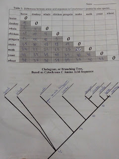
L11: PROTEIN AND EVOLUTION


1. INTRODUCTION


Genes are made of DNA and are inherited from parent to offspring. Some DNA sequences code for mRNA wich, in turn, codes for the amino acid squence of proteins, Cytochrome C is a protein involved in using energy in the cell. Cytochrome C is found in most, if not all, known eukaryotes. Over time, random mutacions in the DNA sequence occur. As a result, the amino acid sequence of Cytochrome C also changes. Cells without usuable C are unlikely to survive.

2. OBJECTIVES


To compare the reladness between organisms by examining the amino acid sequence in the protein, Cytochrom C.

3. PROCEDUCE


 The image is from Ignacio, because I can not find my photocopy.

4. CONCLUSION


As far from each other and are more differences further evolutionarily amino acids. Are grouped by groups of animals.

5. QUESTIONS


1. How many Cytochrom C amino acid sequence differences are there between chickens and tukeys?

0.

2. Make a branking tree, or cladrogram for chikens, penguins and turrrkeys?

chicken- penguin: 0
turkey-penguin: 3

3.a Predict the number of Cytochrom C amino acid sequence differences you would expect to see between.

Horse-Zebra: 1-2

Donkey-Zebra:1-2

b) What other information did you use to make this prediction?

If they can repoduce and the offpring is festile.

5. List three other things used to determine how organisms are related to each other.

Comparing the organs, anatomic prove, embrions,,,

6.  Explain why more closely related organisms have more similar Cytochrome C.

Evolutionarily not so long ago parted hence have not so many mutations.

7. Other data, including other genes, suggest that fugi are more closely related to animals than plants. What are some reasons that the Cytochrome C data suggest that fugi, plants, and animals are equally distantly related?

If you have more than 40, suffered a lot of mutations and can not be compared. 


viernes, 13 de febrero de 2015

L10: PROTEIN DESNATURALITION I

1. INTRODUCTION


Desnaturation is a process in which proteins or nucleic acids lose the quaternary, tertiary and secondary structure that is present in their native state. Desnaturation is the result of the application of some external stress ( heat and pH change) or base, a concentrated inorganic salt or organic solvent.

If proteins in a living cell are denatured, this results is disruption of cell activity and possibly cell death. Denatured proteins can exhibit a wide range of ring characteristics, from loss of solubility to communal aggregation.
This las effect results from the bonding of the hydrophobic proteins to reduce the total area exposed to water. In very few cases denaturation is reversible and proteins can recuperate their native state when the denaturation is reversible and proteins can recuperate their native state when the denaturing factor is removed. This process is called renaturation.

2. OBJECTIVES


- Study the relation between the structure and the function of proteins,

- Understand how temperature, pH snd salinity affect to the protein structure.

3. MATERIAL


- 2X50 mL beaker
- 4 test tubes
- Test tubes rack
- 10 mL pipet
- Knife
- Glass marking pen
- Potato
- Distilled water
- Hydrogen peroxide
- NaCl
- HCl

3. PROCEDUCE


In this experiment we are going to test the catalase activity in different environment situacions. We are going to measure the rate of enzyme activity under various conditions, such as different pH values and temperatures. We will mesuare catalase activity by observing the oxygen gas bubbles when H2O2 is destroyed. If lots of bubbles are produced, it means the reaction is happening and the catalase enzyme is very active,

-  Prepare 30 mL of H2O2 10% in a beaker ( use a pipet).
-  Prepare 20 mL of HCl 10% in a beaker.
-  Prepare 30 mL of NaCl 50 % in a beaker.
-  Peel a fresh potato tuber and put the tissue.
- Label 5 test tubes (1,2,3,4,5).
- Immerse 10 minutes your piece of potato inside HCl and NaOH beaker, and mashed up the potato.
- Add 5 mLH2O2 10% in each test tube.
- With a glass- marking pen mark the height of the height of the bubbles.
- Compare the results of the 5 test tubes.

4. OBSERVATIONS 


- Independent variable: treatment of each potato.
- Dependent variable: the height of the bubbles.
- Experimental Group(s): the rest.
- Control Groups: treatment of each potato.
- Constants: size, amount of H2O2, time...

5. CONCLUSION


Mashed > Raw > NaCl > HCl > Potato boiled.

6. QUESTIONS


1. How did the temperature of potato affect the activity of catalase?

The enzime X of the catalase.

2. How did the change of the Ph of the potato affect the activity of the catalase?

The pH change for acid the catalase no activity while ppH change for a basic the catalase activity.

3. In wich potato treatment was catalase the most active? Why do you think this was?

The mashed up potato , because when we mashed up the potato break the enzyme X.


domingo, 4 de enero de 2015

L9: PROTEIN IDENTIFICATION

1. INTRODUCTION

Biuret's test is a chemical test used for detecting the presence of peptide bonds. A peptide bond can be broken by hydrolysis (the adding of water). In organisms, protein molecules called enzymes facilate the process.
The biuret reaction can be used to assess the concentracion of proteins because peptide bonds occur with the same frequency of amino acid peptide. 
The solution to be tested in treated with a strong base followed by a few with a strong based followed by a few drops of copper (II) sulphate. If the solution turns purple, protein is present.


2. OBJECTIVES


- Identify peptide bonds.
- Compare protein concentracion in differents foods.


3.MATERIAL


- 7 x 250mL beaker
- 6 test tubes
- Test tube rack
- 6 x 10mL Pipet
- Mortar
- Glass marking pen
- Gloves
- Goggles
- Milk
- Soy milk
- Egg
- Yogurt 
- Potato
- Distilled water
- NaOH 20%
- 10 drops of CuSO4


4. PROCEDURE


First of all are going to dilute the protein,

1. Add 100mL of distilled water to each 250mL beaker. Label them with M (milk), S (soy milk) and EW ( egg white), EY (yolk), Y (yogurt) and P (potato).

2. Separate the egg white nd the yolk in another beaker.

3. Smash the potato in a mortar and add some amount of the smashed potato to the P beaker.

Prepare the samples:

4. Add 10 mL of a dispersion of each food ( M,S,EY,EW and Y) to the indicate beaker. Calculate the final concentracion. All the groups will use the same dispersion from the beakers.

5. Prepare 6 test tubes and label M,S,EW,EY,Y and P. Add 2 mL to test tube of the every food disolution of each beaker.

6. Add 2 mL of 20% NaOH dissolution.

7. Shake gently and add 5 drops of CuCO4 in each tube. Allow the mixture to stand for 5 minutes.

8. Note any colour change. Remember that proteins will turn solution pink or purple.

9. Compare the test tubes.


5. OBSERVATIONS


6. QUESTIONS


1. Which food has protein? 
Animal food.

2. Which food has more proteins? Why?
Eggs white, yogurt, potato and milk. Are animal food.

3. Do you find any different between soy and cow milk?
Rice milk, doesn't have protein, however has milk has.

4. Is there any different among milk and yogurt?Why?
Yes. Yogurt has more protein than milk.


martes, 2 de diciembre de 2014

L8: SAPONIFICATION

1. INTRODUCTION

Saponification is a reaction of a fatty acid with a strong base. This reaction forms a salt called soap.

2. OBJECTIVES

- To create a soap.


3. MATERIAL

-2 beakers of 250 mL
-Watch galss
-Spatula
-Stirring rod
-Sosa
-Water
-Oil
-Balance

4.PROCEDURE

First,we have taken a 250 ml beaker and we put 90 mL of water. We put another beaker with 270 mL of oil, and on a watch glass we have mesured 32 grams of sosa. We put the sodium hydroxide into the water  and mixed ( we have tested wich kind of reaction takesplace). Then put the oil very slowly and removed.


5.OBSERVATIONS

We can obtain the soap.

L7: LIPIDS PROPERTIES

1. INTRODUCTION

Lipids are heterogeneous group of compounds synthesized by organisms, that are present in all biological tissues. These compounds are characterized as natural substances that do not mix with water but dissolve in organic solvents, There are several classes of lipids including: fatty acids,waxes,tryacyglycerols, phospholipids,terpens and steroids. C,H and O are the principal elements of lipids althought oxygen content is reduced. Lipids are made in general of long chains of hydrocarbons with relatively little oxygen, they tend to be non-polar, meaning they do not dissolve in polar solvents such as water.


2. OBJECTIVES

- Test the solubility of lipids
- Identify lipids in liquids compounds.
- Understand what are an emulsion and the effect of detergents.


3. MATERIAL

- Test tube rack
- 250ml beaker
- Water
- 6 test tubes
- Cellulose paper
- Dropper
- Scissors
- Glass rod
- Olive oil
- Soap (detergent)
- Milk with different fat content
- Petroleum ether
- Ethanol
- Sudan III

4, PROCEDUCE 

- Solubility of some lipids:

1. Clean and dry three test tubes. Label as W (water), ethanol (E), and PE (ether).
2, Add 3 drops of oleic acid to 3 small test tubes.
3. Add 1ml of water in the first test tube (W).
4. Add 1ml of ethanol in the second test tube (E).
5. Add 1ml of petroleum ether in the third test tube (PE)
6. Shake carefully each test tube and record solubility and observations in my worksheet.

- Lipids identificaion:

A) Translucent mark

1. Cut two pieces (10X10cm) of cellulose paper.
2. Put 1 drop of water in the first squared piece. You will see a translucednt spot. Wait for a while and observe what is happening,
3. Put 1 drop of olive oil in the second squared piece of cellulose paper. You will see a translucent spot,

B) Sudan III dye: Be careful red can stain clothes!!!

1, Take the W test tube of the first experiment and add 2 drops of Sudan III.
2. Prepare four test tubes: 3 with different fat content (M1, M2, M3) and soda (S). Add two drops of sudan III and observe the results.

- Pemanent emulsion:

1. Take a 250ml beaker and put 100 ml of water.
2. Add 1ml of olive oil. With a glass rod stir the mixture vigorously and let it stand a few minutes.
3. Make note of what is happening,
4. Add 2 drops of soap and stir the mixture again. Let it stand for a few minutes and notice the diferences between both mixtures,

5. OBSERVATION

 We have been able to observe...

- In the first experiment we have tested that oil is insoluble in water and soluble in ether. In the second test tube the oil and ethanol didn't mix, the oil formed micelles.

- In the second experiment we tried to stain lipids with sudan III, unfortunatelly the dye didn't stain.

- In the last eperiment we tested that soap forms little micelles with oil, in a water solution. This mixture is called permanent emulsion.


6. QUESTIONS


1. From your observation, which compounds can dissolve lipids?

The ether can dissolve the lipids.

2. Do the oil and water mix? what can you conclude about the polarity of the oil if you know that water is polar?

The oil is not soluble in water and remains in the top of the solution.

3. Why is liquid the olive oil at room temperature? and why not the lard?

Because the oil has a melting point much lower melting.

4. Why does a lipid leave a translucent spot on paper?

Because it contains lipids.

5. Which type of milk contains more lipids? Why?

Whole milk.

6. Did the oil and water mix when you added the soap?

It produces a permanent ebulltion.

7. What did the soap do to the fat?

That can't join again.


8. Can you think about process and locations were compounds like the soap would be important to an animal?

Bile acids.